


Home / Training / Manuals / Atlas of breast cancer early detection / Cases
Atlas of breast cancer early detection
Go back to the list of case studies
.png) Click on the pictures to magnify and display the legends
Click on the pictures to magnify and display the legends
| Case number: | 067 |
| Age: | 53 |
| Clinical presentation: | Postmenopausal woman with average risk of developing breast cancer presented with painful right breast lump. On examination, she was found to have a freely mobile soft to firm lump in the right breast. |
Mammography:
 |  |
| Breast composition: | ACR category b (there are scattered areas of fibroglandular density) | Mammography features: |
| ‣ Location of the lesion: | Right breast, lower outer quadrant at 78 oclock, middle third |
| ‣ Mass: | |
| • Number: | 1 |
| • Size: | 7.0 × 6.0 × 6.7 cm |
| • Shape: | Oval |
| • Margins: | Circumscribed with perilesional halo |
| • Density: | High |
| ‣ Calcifications: | |
| • Typically benign: | None |
| • Suspicious: | None |
| • Distribution: | None |
| ‣ Architectural distortion: | None |
| ‣ Asymmetry: | None |
| ‣ Intramammary node: | None |
| ‣ Skin lesion: | None |
| ‣ Solitary dilated duct: | None |
| ‣ Associated features: | None |
| Breast composition: | ACR category b (there are scattered areas of fibroglandular density) | Mammography features: |
| ‣ Location of the lesion: | Right breast, lower inner quadrant at 5 oclock, middle third |
| ‣ Mass: | |
| • Number: | 1 |
| • Size: | 1.0 × 0.6 cm |
| • Shape: | Oval |
| • Margins: | circumscribed |
| • Density: | Equal |
| ‣ Calcifications: | |
| • Typically benign: | None |
| • Suspicious: | None |
| • Distribution: | None |
| ‣ Architectural distortion: | None |
| ‣ Asymmetry: | None |
| ‣ Intramammary node: | None |
| ‣ Skin lesion: | None |
| ‣ Solitary dilated duct: | None |
| ‣ Associated features: | None |
Ultrasound:
 |  |
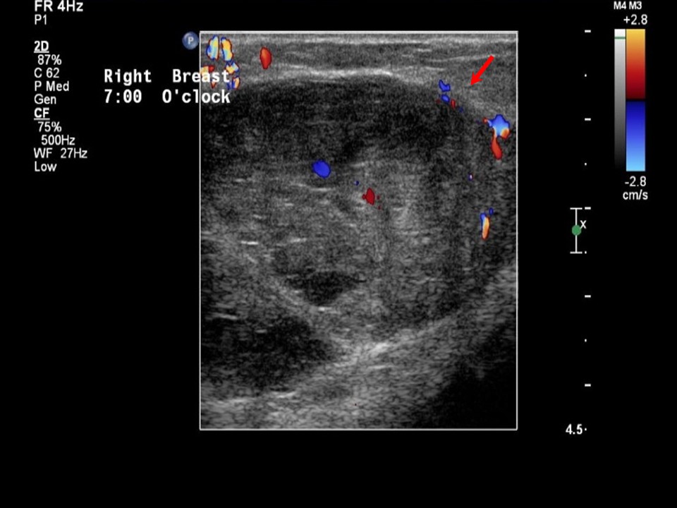 |
| Ultrasound features: Right breast, lower outer quadrant at 7 oclock | |
| ‣ Mass | |
| • Location: | Right breast, lower outer quadrant at 7 oclock |
| • Number: | 1 |
| • Size: | 5.5 × 3.8 cm |
| • Shape: | Oval |
| • Orientation: | Parallel |
| • Margins: | Circumscribed |
| • Echo pattern: | Heteroechoic |
| • Posterior features: | No posterior features |
| ‣ Calcifications: | None |
| ‣ Associated features: | Internal vascularity |
| ‣ Special cases: | None |
BI-RADS:
BI-RADS Category: 4A (low level of suspicion for malignancy)Further assessment:
Further assessment advised: Referral for cytologyCytology:
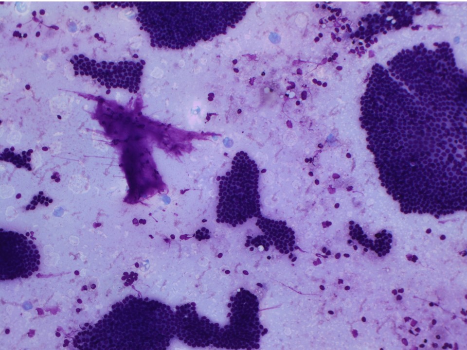 |
| Cytology features: | |
| ‣ Type of sample: | FNAC |
| ‣ Site of biopsy: | |
| • Laterality: | Right |
| • Quadrant: | Outer |
| • Localization technique: | Palpation |
| • Nature of aspirate: | Whitish fluid |
| ‣ Cytological description: | Smears are cellular and show monolayered clusters and sheets of ductal epithelial cells. Myoepithelial cells are seen. A few clusters of apocrine cells are seen. Background shows many foamy cells, macrophages, and haemorrhage |
| ‣ Reporting category: | Benign |
| ‣ Diagnosis: | Benign proliferative breast lesion |
| ‣ Comments: | None |
Histopathology:
Lumpectomy
 |
| Histopathology features: | |
| ‣ Specimen type: | Lumpectomy |
| ‣ Laterality: | Right |
| ‣ Macroscopy: | Breast specimen (6.0 × 5.0 × 3.0 cm) with smooth external surface. Cut surface shows a large cystic area (4.0 × 4.0 × 2.0 cm) with a pedunculated papillary structure. Surrounding breast tissue contains smaller cysts with papillae |
| ‣ Histological type: | Multiple benign papillomas. Sections from the papillary lesions reveal multiple benign papillomas with well-developed fibrovascular cores and broad club-like papillae and arborescent fronds. The broader papillae contain glands. Both the fibrovascular cores and the glandular components are lined by epithelial cells with a prominent myoepithelial layer |
| ‣ Histological grade: | |
| ‣ Mitosis: | |
| ‣ Maximum invasive tumour size: | |
| ‣ Lymph node status: | |
| ‣ Peritumoural lymphovascular invasion: | |
| ‣ DCIS/EIC: | |
| ‣ Margins: | |
| ‣ Pathological stage: | |
| ‣ Biomarkers: | |
| ‣ Comments: | Negative for malignancy |
Case summary:
| Postmenopausal woman presented with painful right breast lump. Diagnosed as mass of suspicious morphology, BI-RADS 4A on imaging, as benign proliferative lesion on cytology, and as multiple benign papillomas on histopathology. |
Learning points:
|